Author: Abdullah Shoaib * 1, Mathew Stokes MD 1
Author Affiliation:
1 Department of Pediatrics, Neurology and Neuroscience at University of Texas Southwestern Medical Center, Dallas, Texas, USA
Competing Interests: The author/s declare no competing interests.
The use of bupivacaine and other local anesthetics to perform peripheral nerve blocks is a mainstay in the management of refractory headaches and migraines. In this case report, a patient presented with transient facial nerve palsy shortly after receiving occipital nerve blocks for headaches.
Patient’s symptoms self-resolved, and her symptoms were thought to be due to displacement and spreading of bupivacaine to the facial nerve. The spread of bupivacaine can be facilitated by tracking across fascial planes or nerve sheaths. Similar side effects have been seen in dental anesthesia, but there has only been one other report of such a presentation associated with occipital nerve blocks, and none in pediatric patients. Physicians should be aware of this rare complication with peripheral nerve blocks.
Introduction
Nerve blocks with medications such as bupivacaine, and other anesthetics is one of the treatment options of intractable headaches. While many patients respond well to nerve blocks and adverse events are rare, this case report details a pediatric patient who developed transient unilateral face weakness shortly after treatment with peripheral nerve blocks. This appears to be a rare occurrence, and there is little reporting of this complication in the current literature, though similar events have also been seen in dental anesthesia cases.
Case Report
A 16-year-old girl with a history of long-standing headaches which began after two concussions (3 years and 1 year prior to the visit), was seen for evaluation and treatment of headaches. Patient’s headaches were holocephalic, worst across her forehead and at base of skull. She described the pain as throbbing and squeezing, though it was occasionally sharp. She had daily, constant pain since her second injury. She rated the pain as a 7 on a 10-point scale. She suffered from migrainous features; e.g., photophobia, phonophobia, as well as nausea. She stated that certain positions (such as looking down) made her pain worse, and that the headaches got worse towards the evening.
Patient had tried physical therapy, treatment by chiropractor, amitriptyline, and topiramate without any significant improvement in her symptoms. Amitriptyline was discontinued due to lack of efficacy at maximal dosage, and topiramate was discontinued due to side effects (appetite suppression, paresthesias). At the time of the visit, patient’s migraine regimen consisted of rizatripran for abortive migraine therapy, as well as multiple PRN agents including acetaminophen, NSAIDs, and topical diclofenac gel without significant improvement in her symptoms. Neurological exam was remarkable only for tenderness over the bilateral greater and lesser occipital nerves, temporoauricular nerves, and supraorbital nerves.
Patient was assessed and diagnosed with chronic migraines. Given nerve tenderness on exam, she was scheduled for nerve blocks and trigger point injections a month after initial consultation. On the day of the nerve blocks, patient endorsed feeling anxious in anticipation of the procedure. Bilateral greater occipital nerve blocks were done first, using 2 mL of 0.5% bupivacaine on each side. She then received bilateral supraorbital nerve blocks with 0.6 mL of 0.5% bupivacaine on each side and bilateral temporoauricular nerve blocks with 0.7 mL of 0.5% bupivacaine on each side. Finally, bilateral lesser occipital nerve blocks were done due to ongoing tenderness in area of the lesser occipital nerve, with 0.7 mL of 0.5% bupivacaine on each side. Prior to each injection, needle was aspirated to ensure physician was not located in vasculature During the final nerve block, the left lesser occipital nerve block, patient developed lightheadedness and stated that she “[felt] like I’m going to pass out”. The procedure was completed as the symptoms developed. Shortly thereafter, she passed out and was laid on the bed on her left side and had brief convulsive movements lasting 3-5 seconds. She regained consciousness after this with immediate return to baseline, with no post-ictal period. Patient was allowed to rest and orally rehydrate for approximately 15 minutes and monitored. During this time, patient reported having immense improvement in her headaches after peripheral nerve blocks and expressed that she wished to continue with trigger point injections.
After discussion with patient and her parents about the risks and benefits of proceeding further, decision was made to proceed with trigger point injections using 0.2 mL of 0.5% bupivacaine in bilateral cervical paraspinal muscles (2 on each side), bilateral thoracic paraspinal muscles (1 on each side), bilateral trapezius (1 on each side), and bilateral splenius capitis (1 on each side). The patient was monitored for return of symptoms with these injections and tolerated them with no issues. The total volume of bupivacaine used in the nerve blocks was 8 mL, and 2 mL were used in trigger point injections.
After completion of nerve blocks and trigger point injections, patient’s headache was completely resolved (0 on a 10-point scale).
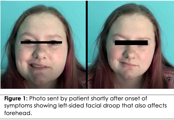
Approximately one hour after the procedure, the patient’s mother contacted due to new onset of left sided facial weakness (see Figure 1). The family provided photos to the clinic at the time of the call, and these were reviewed. Given that the patient had a peripheral pattern of weakness, acute imaging was not obtained. A course of steroids was prescribed to start if the symptoms did not resolve. Approximately 4-5 hours after the injections, the patient’s symptoms resolved completely, so steroids were not initiated. A routine brain MRI was done roughly 2 months after this event due to persistent and worsening headaches and revealed no evidence of infarction, prior infection, or any other abnormalities.

Discussion
There are two events and teaching points seen in this patient case—the patient’s syncopal event and their subsequent facial nerve palsy. There was concern that patient’s acute loss of consciousness and lightheadedness following use of local anesthetic, may have been due to local anesthetic systemic toxicity (LAST)1. When local anesthetic is injected, it can diffuse into the bloodstream and cause systemic side effects 1, 2. Using high doses of local anesthetics, causing higher concentration gradients, and injecting local anesthetic intravascularly increase risk of local anesthetic causing systemic side effects 1. The primary mechanism of action of local anesthetic is blockade of voltage-gated sodium channels on the membrane 1, 2. When used in peripheral nerve blocks, blockade of sodium channels blocks generation and propagation of action potential in peripheral nerves, thereby reducing propagation of pain from peripheral nerves. However, if high enough concentrations of local anesthetic are attained in cardiac tissue, the sodium channel blockade can cause catastrophic arrhythmias 1. Similarly, in high concentrations, lidocaine activity on sodium channels in the brain can cause seizures 1.
Typical manifestations of LAST are dysphoria, dizziness, light-headedness, tinnitus, or drowsiness, and can then progress to seizure, cardiac arrhythmias, hypotension, and asystole 1, 2. Treatment of LAST involves stopping the inciting medication, providing oxygen and ventilation if necessary, controlling seizure with benzodiazepines, and ACLS 1. In addition, use of intravenous lipid emulsion has been shown to help by allowing the anesthetic to diffuse into the lipid and then be transported away from cardiac tissue 1. Physicians can minimize risk of LAST by aspirating the needle prior to injecting local anesthetic to ensure they are not injecting into vasculature, and by using lower doses of local anesthetic 1, 2. In addition, using ultrasound to ensure that the needle is not close to vascular structures can also help reduce risk of LAST 2.
Due to the patient’s prodrome of feeling lightheaded, brief duration of loss of consciousness (only a few seconds) with associated limb-stiffening, and quick return to baseline with no post-ictal state, the index of suspicion for seizure was low. Her episode was felt to be more consistent with a vasovagal etiology. Patient’s anxiety surrounding the procedure was also likely to have contributed to patient’s vasovagal syncopal episode. It is possible that patient’s prodromal symptoms of lightheadedness could represent a mild form of LAST. It is possible that bupivacaine was injected intravascularly, though this risk was minimized by ensuring no blood was aspirated by the needle prior to injection. In addition, it is possible that local anesthetic diffused into vasculature, causing systemic toxicity. Overall, LAST seems less likely, given the fact that doses of bupivacaine administered were much lower than those associated with LAST (40 mg bupivacaine were used in total for all nerve blocks vs. doses of greater than 150 mg bupivacaine are typically associated with LAST) and the patient’s rapid recovery back to baseline with no further symptoms.
Understanding the anatomy of the facial nerve is necessary to fully appreciate and understand the patient’s unexpected facial nerve palsy. The motor and sensory roots that eventually join to become the facial nerve originate from the pons. These two roots travel through internal acoustic meatus, which is in close proximity to the inner ear. The roots exit the internal acoustic meatus and enter the facial canal, where these two roots join together to become the facial nerve. Various branches of the facial nerve come off the facial nerve while intracranially in the facial canal, such as the chorda tympani, which is involved in taste, and the nerve to the stapedius muscles in the middle ear. The facial nerve exits the facial canal via the stylomastoid foramen, close to the temporoauricular nerve, where it then begins to travel extracranially, close to the posterior ear. One of the first extracranial branches of the facial nerve is the posterior auricular nerve, which lies close to but laterally to the lesser occipital nerve. Branches of the facial nerve involved in facial expression also branch off close to the origin of the posterior auricular nerve3. Of note, autopsy studies have also revealed considerable variation in the size and course of the facial nerve 4.
A literature review found only one prior report of a similar case of facial nerve palsy 4. In this case report, a 24-year-old patient with a history of migraines underwent left greater and lesser occipital nerve blocks with bupivacaine and triamcinolone. Within minutes of completion of these nerve blocks, patient reported anxiety and tingling and paresthesia to left upper lip, which subsequently spread to her complete left face, and was noted to have water dribbling out of her left lip when she tried to take a drink. She had noticeable left sided facial weakness, with flattening of left nasolabial fold, drooping of left eyebrow and forehead, and decreased blinking on left. Patient had an MRI of the brain with and without contrast to assess for any possible hematoma or fluid collection causing impingement of facial nerve, which was negative. The patient’s symptoms resolved within 5 hours of onset of symptoms 4.
The authors of this case report felt that this transient facial paralysis was due to bupivacaine, due to the temporal relationship between the nerve blocks and facial paralysis, as well the fact that the duration of symptoms was consistent with the pharmacodynamics of bupivacaine. The authors posited that infiltration of bupivacaine into communicating tissue planes or inadvertent injection into a neural sheath near the lesser occipital nerve may have led to spread to the left facial nerve, causing the patient’s symptoms 4. The authors note that that the posterior auricular nerve lies in close proximity to the lesser occipital nerve, and that it is possible that the bupivacaine may have been injected into the nerve sheath or fascial tissue of the posterior auricular nerve, where it then tracked back to the main facial nerve trunk 4.
While there is limited reporting of facial nerve palsy following nerve blocks for migraines, other nerve blocks have been associated with this. Many case reports detail transient facial palsies after dental nerve blocks, particularly, inferior alveolar nerve blocks 5, 6, 7. These patients developed ipsilateral facial nerve palsy following nerve blocks—in one case 5 symptom onset was within a few hours, and in another 6, symptoms began over 24 hours after the procedure. In the third of these studies, the patient’s symptoms were apparent a few minutes after the procedure, and completely resolved within 5 hours 7. The authors of these papers 5, 6, 7 classified facial nerve palsies after inferior alveolar nerve blocks into two categories: immediate, wherein symptoms begin minutes after injection of anesthetic and resolve quickly, and delayed, wherein symptoms begin hours after injection of anesthetic and linger for weeks to months. They propose that the immediate paralysis could be due to local infiltration of anesthetic solution via local tissue or fascial planes, ultimately affecting the facial nerve, and that the degree of symptoms may be influenced by any aberrance in the course or position of the trunk of the facial nerve or any subsequent branches 5, 6, 7. They contrast that with those facial nerve palsies that have delayed onset but have lingering symptoms, whose exact pathophysiology is unclear, but could be due to the procedure triggering viral reactivation leading to a Bell’s palsy, or ischemic injury to the facial nerve due to either mechanical trauma due to prolonged instrumentation or a reflex vasospasm due to stimulation of the sympathetic plexus of the external carotid artery 5, 6, 7. Of note, in two of these studies, mepivacaine and articaine were used instead of bupivacaine 5, 6.
In the case of our patient, she contacted clinic when symptoms had peaked about an hour after the injection and likely had developed symptoms gradually over that time. She also had rapid recovery. It seems that her symptoms were likely due to infiltration of the anesthetic into her facial nerve via fascial planes.
The most likely explanation is that patient’s syncopal episode after the lesser occipital nerve block caused some displacement of the bupivacaine into a tissue plane that could facilitate transfer to the facial nerve. Among the nerve blocks performed, the location of the lesser occipital nerve block, close to the posterior auricular branch of the facial nerve 3, and temporoauricular nerve block, close to the extracranial exit of the facial nerve 3, are among the closest to the proximal facial nerve and thus, one of these was most likely to cause facial nerve palsy. It is possible that after leaving the clinic, the patient may have positioned herself in such a way that could have facilitated transfer of bupivacaine to the facial nerve, which could explain the minor delay in onset of symptoms, as well as the rapid recovery. Of these two nerve blocks, it is difficult to determine which one may have caused the facial palsy. Once the bupivacaine came in contact with left facial nerve, it blocked sodium channels and inhibited action potential, causing paralysis of facial nerve.
Per our literature review, this is the first report of transient facial nerve paralysis after nerve blocks for chronic migraines in a pediatric patient, and only the second detailing similar symptoms in patients receiving nerve blocks for chronic migraines. Nerve blocks are not uncommonly used for intractable migraines, and transient facial nerve palsies are a rare complication that clinicians should be aware of. The timing of onset of symptoms can shed some light on the likely cause of the facial nerve palsy and can be helpful in predicting the trajectory of symptoms. In addition, physicians should also be aware of possible systemic toxicity due to local anesthetic use and should be judicious when using local anesthesia and may consider using imaging to ensure anesthetic is not being injected intravascularly.
References
- Gitman M, Fettiplace MR, Weinberg GL, et al. Local Anesthetic Systemic Toxicity: A Narrative Literature Review and Clinical Update on Prevention, Diagnosis, and Management. Plast Reconstr Surg. 2019 Sep;144(3):783-795. PubMed CrossRef
- Dickerson DM, Apfelbaum JL. Local anesthetic systemic toxicity. Aesthet Surg J. 2014 Sep;34(7):1111-1119. PubMed CrossRef
- Augur AMR, Dalley AF. Grant’s Atlas of Anatomy 14th Philadelphia, PA: Wolters Kluwer; 2017. Chapter 9, Overview of Cranial Nerves; p.783-816.
- Strauss L, Loder E, Rizzoli P. Transient facial nerve palsy after occipital nerve block: a case report. 2014 Nov-Dec;54(10):1651-1655. PubMed CrossRef
- Chevalier V, Arbab-Chirani R, Tea SH, Roux M. Facial palsy after inferior alveolar nerve block: case report and review of the literature. Int J Oral Maxillofac Surg. 2010 Nov;39(11):1139-1142. PubMed CrossRef
- Tzermpos FH, Cocos A, Kleftogiannis M, et al. Transient delayed facial nerve palsy after inferior alveolar nerve block anesthesia. Anesth Prog. 2012;59(1):22-27. PubMed CrossRef
- Jenyon T, Panthagani J, Green D. Transient facial nerve palsy following dental local anaesthesia. BMJ Case Rep. 2020 Sep 7;13(9):e234753. PubMed CrossRef
Disclosures
Consent/Permissions: Patient consent was received to report the case.
Funding: No sources of funding.
Conflicts of interest: In compliance with the ICMJE uniform disclosure form, all authors declare the following: Payment/services info: All authors have declared that no financial support was received from any organization for the submitted work. Financial relationships: All authors have declared that they have no financial relationships at present or within the previous three years with any organizations that might have an interest in the submitted work. Other relationships: All authors have declared that there are no other relationships or activities that could appear to have influenced the submitted work.
