Author: Amir Soheil Tolebeyan* MD 1
Author Affiliation:
1 Assistant Professor, Headache and Facial Pain, Department of Neurology, Tufts Medical Center, Tufts University School of Medicine
Competing Interests: The author/s declare no competing interests.
Issue: 09.02
DOI: 10.30756/ahmj.2022.09.02
Received: Oct 5, 2022
Revised: Nov 21, 2022
Accepted: Dec 31, 2022
Published: Jan 13, 2023
Recommended Citation: Tolebeyan AS. Post-Surgical CSF leak with Inconclusive Diagnostic Studies: A Case Report. Ann Head Med. 2022;09:02. DOI: 10.30756/ahmj.2022.09.02
Objectives: To report a case with post-surgical intracranial hypotension and inconclusive diagnostic findings.
Background: The diagnosis of spontaneous intracranial hypotension is based on positive neuroimaging findings and/or low CSF pressure on spinal tap. Despite this conventional definition, cases with normal CSF pressure have also been reported.
Results: This report explains a patient with post-surgical CSF leak, normal CSF opening pressure, and unremarkable imaging studies that responded well to the surgical repair.
Conclusion: Our case challenges the conventional diagnostic criteria of spontaneous intracranial hypotension. We suggest that clinical judgment should be considered in regard to the diagnosis of spontaneous intracranial hypotension.
Introduction
Spontaneous intracranial hypotension (SIH) is a relatively uncommon diagnosis with estimated prevalence of up to 5/100000 of general population described for the first time in 1938 1, 2. SIH diagnosis is made based on positive neuro imaging findings and/or low CSF pressure on lumbar puncture 3. However, despite the conventional definition of SIH, some investigators have reported normal range of CSF pressure among patients with diagnosis of SIH 4-6.
Case Report
A 48-year-old female presents with new onset headache following L4-L5 revision laminectomy/discectomy for left L5 radiculopathy with left lower extremity weakness. The patient has a past medical history for episodic migraine.
The patient started to experience excruciating headaches a few hours following the surgery. She described the headaches as 4/10-8/10, dull, constant bifronto-temporal pain with jaw pressure and neck stiffness. The patient reported 4/10 constant daily headaches with 6-8/10 exacerbations a couple of times a week. Other accompanying symptoms were photophobia, phonophobia, nausea, and occasional vomiting. The headaches were aggravated by physical activity, bright light, noises, and upright position. Most of her headaches improved within 20 minutes after lying down.
Medical history included anxiety and depression, type II diabetes mellitus, low back pain, and menstrual migraine. She was on cyanocobalamin, medical marijuana, diazepam, sertraline, and metformin. Her neurologic examination was normal.
The patient was given zolmitriptan 5 mg tablets and ibuprofen, acetaminophen/butalbital/caffeine for the acute attacks, which provided pain relief, but not a pain freedom.
CT lumbar spine a couple of weeks following the surgery was suggestive of postsurgical laminectomy changes at L4 and L5 with opacity traversing the laminectomy defects, stenting posteriorly into the interspinous region, which may represent expected postoperative changes.
CT scan of the head and CTA of the head and neck without IV contrast and MRI of the brain with and without IV contrast performed three months after surgery did not show any abnormal findings.
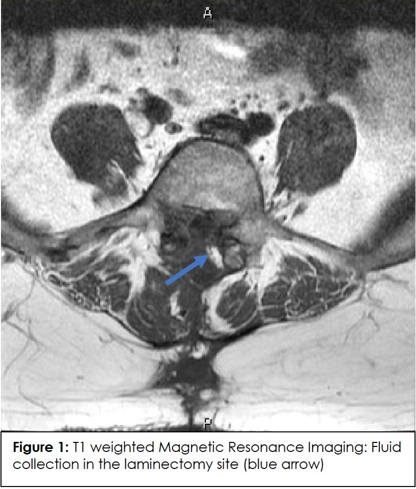
The patient was evaluated by a headache specialist for the first time about three months post-surgery, and an MRI of the cervical, thoracic and lumbar spine was subsequently requested. The MRI of the spine was indicative of some degenerative changes throughout the spinal column and fluid collection in the laminectomy site extending to the posterior superficial soft tissues (Figure 1).

A CT myelography was performed about six months following the MRI spine, indicative of postsurgical changes at the L4-S1 level without evidence of contrast extravasation into the soft tissues or the epidural space. The opening pressure was 18.5 cm H2O on lumbar puncture.
The patient had a follow-up four days after the CT myelogram.
The patient remained symptomatic and resistant to the standard pharmacotherapy. The patient had tried multiple medications, including zolmitriptan, intravenous fluids, NSAIDs, and magnesium oxide, with no consistent relief. She eventually underwent an epidural blood patching with 45 CCs autologous blood at the level of L2-L3 approximately ten months after the surgery. It did not provide any headache relief.
Despite inconclusive imaging studies, the CSF leak was still suspected, given the phenotype and timeframe of headaches that started following the surgery. The patient underwent surgical repair of dural CSF leak and pseudo meningocele about 15 months after the initiation of headaches. The patient’s symptoms improved dramatically following the procedure, with complete resolution of neck stiffness and positional headaches a few days later.
Discussion
According to the International Classification of Headache Disorders, third edition (ICHD3), spontaneous intracranial hypotension is defined as a headache in temporal relation to the low CSF pressure and either low CSF pressure (<60 mmH2O) and/or positive imaging findings suggestive of low CSF pressure or volume 3. However, it seems that this definition is not comprehensive enough to encompass all cases with SIH.
Despite the conventional definition of SIH, which includes low CSF pressure, some investigators have reported a normal range of CSF pressure among patients with the diagnosis of SIH 4-6. These findings change the concept of low CSF pressure as the primary etiology of spontaneous intracranial hypotension.
Cun Li and colleagues analyzed retrospective data from 40 patients with confirmed SIH 5. All patients had signs of orthostatic headaches. MRI of the brain with gadolinium was performed on 24 patients. Of those, pachymeningeal enhancement was seen in 23 patients (95.83%), 5 patients (20.8%) had pituitary hyperemia, the subdural fluid collection was seen in 4 cases (16.7%), brain sagging in 3 cases (12.5%), and venous engorgement was seen in 1 case (4.1%). A lumbar puncture was performed in all 40 patients. Cerebrospinal fluid pressure was <60 mmH2O in 37 patients (92.5%) 5.
Kranz and coworkers performed a retrospective review on 106 patients with SIH and measured the opening CSF pressure on lumbar puncture prior to treatment 6. 82% showed evidence of dural enhancement, 60% brain sagging on gadolinium-enhanced MRI. However, 9% of patients had negative brain imaging. In the case of CSF opening pressure, only 34% had CSF opening pressure (CSF OP) ≤ 60 mm H2O, while CSF OP was between 6 and 20 cm H2O in 61% of patients, and 5% had PCSF > 20 cm H2O 6.
Conclusion
Our case study challenges the conventional diagnostic criteria of SIH in two ways; First, not all cases with SIH present with positive findings on imaging studies, and second, low CSF pressure might not be present in all SIH patients. This case’s clinical presentation, and diagnostic findings along with dramatic response to treatment, emphasizes the fact that not all patients with SIH fulfill the ICHD-3 criteria of spontaneous intracranial hypotension and clinical judgement should be considered strongly in regards with clinical presentation, especially with cases with post operational headaches or those with accelerated headaches happening post spinal procedures.
References
- Couch JR. Spontaneous intracranial hypotension: the syndrome and its complications. Curr Treat Options Neurol. Jan 2008;10(1):3-11. PubMed PMID: 18325294. doi:10.1007/s11940-008-0001-5
- Renowden SA, Gregory R, Hyman N, Hilton-Jones D. Spontaneous intracranial hypotension. J Neurol Neurosurg Psychiatry. Nov 1995;59(5):511-5. PubMed PMID: 8530936; PubMed Central PMCID: PMCPMC1073714. doi:10.1136/jnnp.59.5.511
- 7.2 Headache attributed to low cerebrospinal fluid (CSF) pressure. IHS Classification ICHD-3. Accessed 1/3/23, 2023. https://ichd-3.org/7-headache-attributed-to-non-vascular-intracranial-disorder/7-2-headache-attributed-to-low-cerebrospinal-fluid-pressure/
- Mokri B, Hunter SF, Atkinson JL, Piepgras DG. Orthostatic headaches caused by CSF leak but with normal CSF pressures. Neurology. Sep 1998;51(3):786-90. PubMed PMID: 9748027. doi:10.1212/wnl.51.3.786
- Li C, Raza HK, Chansysouphanthong T, Zu J, Cui G. A clinical analysis on 40 cases of spontaneous intracranial hypotension syndrome. Somatosens Mot Res. Mar 2019;36(1):24-30. PubMed PMID: 30870079. doi:10.1080/08990220.2019.1566122
- Kranz PG, Tanpitukpongse TP, Choudhury KR, Amrhein TJ, Gray L. How common is normal cerebrospinal fluid pressure in spontaneous intracranial hypotension? Cephalalgia. Nov 2016;36(13):1209-1217. PubMed PMID: 26682575. doi:10.1177/0333102415623071
Declarations/Disclosures
Consent/Permission/Ethics Approval: A single case with no identifiable data. There are no potentially identifiable human images or data presented in the manuscript. None of the patient’s information included is identifiable to any specific individual.
Conflicts of interest: In compliance with the ICMJE uniform disclosure form, author declares the following:
Payment/services info: Author has declared that no financial support was received from any organization for the submitted work.
Financial relationships: Author have declared that they have no financial relationships at present or within the previous three years with any organizations that might have an interest in the submitted work.
Other relationships: Author have declared that there are no other relationships or activities that could appear to have influenced the submitted work.
